Figure 4.
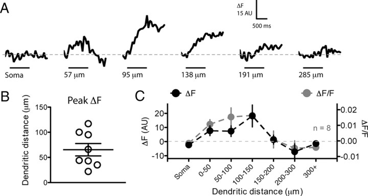
The initial 150 μm of the dendrite has a high sodium channel density. GnRH neurons were whole-cell loaded with 500 μm of the Na+-sensitive dye CoroNa Green. After 15–20 min loading, neurons were induced to spike 10 times at 10 Hz with step depolarizations. Changes in CoroNa fluorescence were subsequently measured with frame scans on the soma and different regions of the dendrite. A shows ΔF traces from one neuron at six different imaging locations. The black bar under traces indicates the period of spiking at the soma. Each trace is the average of 5–10 trials. In this neuron, the largest Na+ transient was observed at a site 95 μm from the soma. B, The site of the largest ΔF Na+ transient is plotted for eight different neurons. C, The average ΔF (black) and ΔF/F (gray) per section of dendrite for eight neurons shows large Na+ transients in the first 150 μm of dendrite but little or no transients at distances >150 μm. For the graph in C, only the largest response per region of dendrite is plotted for each neuron.

