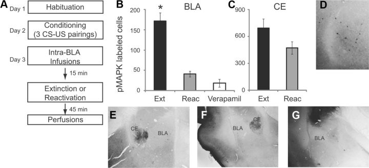
Figure 2.
Extinction training induces pMAPK in the BLA which is blocked by intra-BLA infusions of verapamil. A, Schematic of behavioral protocol. B, Number of pMAPK-labeled cells in the BLA after ACSF infusion followed by extinction (Ext) or reactivation (Reac) or verapamil infusion followed by extinction (Verapamil). C, Number of pMAPK-labeled cells in the CE after ACSF infusion followed by extinction (Ext) or reactivation (Reac). D–G, Example images showing pMAPK-labeled cells at high magnification in the BLA (D), following ACSF extinction (E), ACSF reactivation (F) and Verapamil extinction (G).

