
Figure 4.
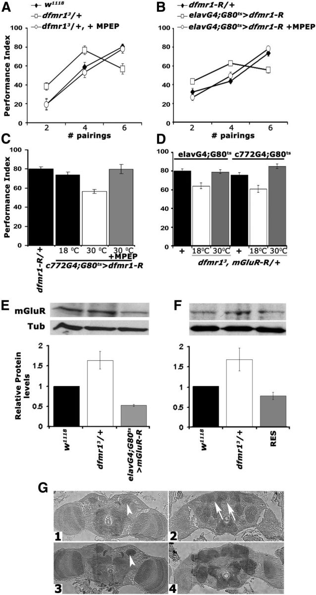
Genetic and pharmacological inhibition of DmGluRA restores the learning deficit of dfmr1 mutants. Mean PIs and their SEMs (PI ± SEM) are shown for all experiments. Where appropriate, animals were heterozygous for transgenes, Gal4 and Gal80ts. All animals were trained and tested at 25°C, regardless of the temperatures they were maintained before training to manipulate transgene expression as indicated. A, MPEP administration to dfmr13/+ reversed the aberrant learning of the heterozygotes (open squares) after two, four, and six pairings to control levels (filled diamonds). n ≥ 10. ANOVA indicated significant differences (p < 0.001) resolved by subsequent Dunnett's tests, which revealed that for two, four, and six pairings the performance of treated animals (open circles) was indistinguishable from controls (p = 0.55, p = 0.51, and p = 0.96, respectively), whereas that of untreated ones remained significantly different at all pairings (p < 0.0001). B, Pharmacological rescue (open circles) of learning in animals with pan-neuronal dfmr1 abrogation (open squares) to control levels (filled diamonds) of performance after training with two, four, and six pairings. The differences suggested by ANOVA (p < 0.001) were examined by Dunnett's tests and demonstrated that treated animals (open circles) and controls (filled diamonds) did not perform differently after two, four, and six pairings (p = 0.21, p = 0.57, and p = 0.23, respectively), whereas untreated animals performed significantly different from both regardless of pairings (p < 0.0001). C, Conditional abrogation (30°C) of dfmr1 in the MBs with c772 impairs learning (open bar) and can be rescued by MPEP (gray bar) to control levels (black bars). n = 10. The performance of animals carrying both transgenes but not expressing UAS-dfmr1-R (18°C) was used to compare that of experimental animals to (Dunnett's tests), after positive initial ANOVA (p < 0.0001). Transgene induction resulted in significant differences with control performance (p < 0.0001), but not after MPEP treatment (p = 0.54). D, Genetic rescue of the learning deficit in dfmr13/+ by pan-neuronal (left three bars) and MB limited conditional attenuation of DmGluRA. Heterozygotes for the Gal4 driver, Gal80ts with the w1118-derived chromosomes were used as controls (black bars) for Dunnett's tests in both cases. n ≥ 10. Maintaining the mGluR-R transgene inactive either pan-neuronally, or in the MBs of dfmr13 heterozygotes resulted in deficient six pairing learning (p < 0.0001). However, abrogation of DmGluRA pan-neuronally, or specifically in the MBs, resulted in performances similar to those of controls (p = 0.23 and p = 0.02, respectively). E, Semiquantitative Western blot analysis of mGluRA levels in adult head lysates from dfmr13/+ flies with pan-neuronal abrogation of DmGluRA and controls. A representative blot of four is shown above, and the quantification below includes all replicates. mGluRA levels relative to the Tubulin loading control were arbitrarily set to 1 for the w1118 controls, and the mean relative levels ± SEM in the experimental animals were estimated and plotted. Levels in the experimental animals were significantly different from those in controls (Dunnett's, p < 0.001). F, A representative semiquantitative Western blot of mGluRA levels in adult head lysates from w1118 controls, dfmr13/+, and their sibling dfmr13/+ heterozygotes carrying a single copy of the dfmr1 genomic transgene (RES). mGluRA levels relative to the Tubulin loading control were arbitrarily set to 1 for the w1118 controls and the mean relative levels ± SEM in the experimental animals were estimated and plotted. mGluRA levels in the experimental animals were not significantly different from those in controls, but were significantly different from those in dfmr13/+ (Dunnett's, p < 0.001). G, Five micrometer frontal paraffin sections from adult heads of w1118 (G1, G2) and dfmr13/+ (G3, G4) flies challenged with α-mGluRA. Sections are shown at the level of the MB calyces (G1, G3, arrowheads) in the posterior of the head and in the mid-anterior of the head (G2, G4). The arrows in G2 point to the MB axonal projections (lobes), which are not stained in agreement with prior reports (Devaud et al., 2008). DmGluRA elevation is apparent in dfmr13/+ brains, which were processed on the same histological slide as controls.