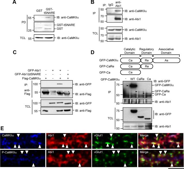
Figure 1.
Abi1 tSNARE domain interacts with CaMKIIα catalytic domain. A, Mouse brain lysates were incubated with purified GST or GST-tSNARE protein, and the bound proteins to GST fusion proteins were pulled-down (PD) by glutathione Sepharose beads and immunoblotted (IB) with anti-CaMKIIα antibodies. GST fusion proteins were visualized by Coomassie Blue staining. B, Lysates from 14 DIV rat cortical neurons were immunoprecipitated (IP) with control polyclonal IgGs or anti-Abi1 antibodies (anti-Abi1) and immunoblotted with anti-CaMKIIα antibodies and anti-Abi1 antibodies. C, Lysates from HeLa cells expressing Flag-CaMKIIα and either GFP-Abi1 or GFP-Abi1ΔtSNARE were immunoprecipitated with anti-Abi1 antibodies and immunoblotted with anti-GFP antibodies and anti-Flag antibodies. D, Lysates from HeLa cells expressing GFP-CaMKIIα (wild-type), GFP-CaRe (catalytic domain and regulatory domain), or GFP-Ca (catalytic domain) were immunoprecipitated with anti-Abi1 antibodies and immunoblotted with anti-GFP antibodies and anti-Abi1 antibodies. Arrowheads (▶) indicate coimmunoprecipitated CaMKIIα proteins with Abi1. Asterisks in C and D indicate heavy chains of IgGs in the immunoprecipitates. E, Immunocytochemistry of 14 DIV rat hippocampal neurons in the top panels shows partial colocalization of the endogenous Abi1 (red) and CaMKIIα (blue) at synapses visualized with anti-vGlut1 antibodies (green). Bottom panels show P-CaMKIIα (blue) localization with Abi1 (red) and vGlut1 (green). White arrowheads indicate regions of vGlut1 staining in each panel. Scale bar, 10 μm. TCL, Total cell lysate.

