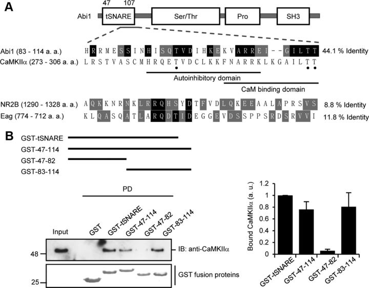
Figure 2.
Abi1 tSNARE has high homology to CaMKIIα regulatory domain and mediates Abi1 binding to CaMKIIα. A, Schematic representation of Abi1 and sequence alignment of Abi1, NR2B, and Eag channel to CaMKIIα. P-CaMKIIα sites are indicated with black dot. Amino acids conserved between CaMKIIα and other proteins are highlighted in black, and conservative substitutions are in gray. B, Top, Schematic diagram of GST-tSNARE deletion mutants. Middle, In vitro GST pull-down (PD) assay. Purified rat brain CaMKII were incubated with indicated GST fusion proteins and analyzed for binding to CaMKII by immunoblotting (IB). GST fusion proteins were visualized by Coomassie Blue staining. Bottom, Results from three independent experiments were quantified, normalized to amount of GST fusion proteins, and expressed in arbitrary units (a. u.). Pro, Proline-rich domain.

