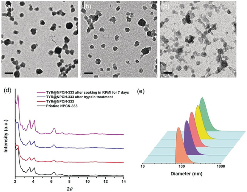
Figure 2.
Structural characterization of NPCN-333. TEM images of NPCN-333 (a), TYR@NPCN-333 (b), and TYR@NPCN-333 after soaking in RPMI-1640 for 7 days (c). Scale bar: 200 nm. d) Powder X-ray diffraction (PXRD) patterns of pristine NPCN-333 (black), TYR@NPCN-333 (red), TYR@NPCN-333 after trypsin treatment (blue), and TYR@NPCN-333 after soaking in RPMI 1640 media for 7 days (magenta). e) Particle-size distribution measured by dynamic light scattering (DLS): (from front to back) pristine NPCN-333, TYR@NPCN-333, TYR@NPCN-333 after trypsin treatment, and TYR@NPCN-333 after soaking in RPMI 1640 media for 0 and 7 days.

