Figure 3.

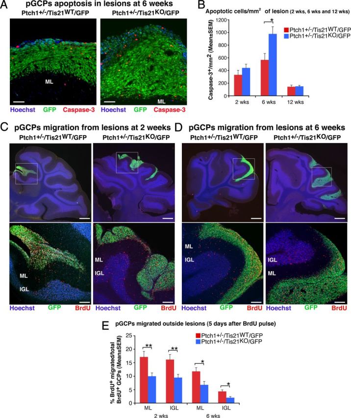
Ablation of Tis21 in Patched1 heterozygous mice increases the apoptosis and impairs the migration from lesions of preneoplastic GCPs. A, Representative confocal images of apoptotic GCPs, identified as cleaved Caspase-3-positive cells, in a diffuse hyperplasia lesion in Patched1 heterozygous mice, either Tis21 wild-type (Ptch1+/−/Tis21WT/GFP) or Tis21-null (Ptch1+/−/Tis21KO/GFP) at 6 weeks of age. Preneoplastic GCPs in the lesion are GFP+; the ML and its boundaries are visualized by Hoechst 33258 staining. Scale bar, 50 μm. B, Apoptotic GCPs per lesion area were quantified as mean ± SEM/mm2 of cleaved Caspase-3+ cells in the whole area of the lesion (defined by the presence of GFP+ cells), on at least five nonadjacent sagittal sections for each lesion. The number of mice analyzed is indicated in Figure 2E. C, D, Representative confocal images of GCPs migrating outside the lesions, identified as BrdU+ cells (red), in Patched1 heterozygous mice either Tis21 wild-type (Ptch1+/−/Tis21WT/GFP) or Tis21-null (Ptch1+/−/Tis21KO/GFP) at 2 or 6 weeks of age. Sections are counterstained with Hoechst 33258 to visualize the ML or the IGL. Lesions in boxed area of top panels (scale bars, 410 μm) are shown at higher magnification in panels below (scale bars, 100 μm). E, GCPs migrating from lesions were quantified as mean ± SEM percentage ratio of BrdU+ cells present within the ML or the IGL area neighboring each lesion 5 d after the BrdU pulse, to the total number of BrdU+ cells in lesion, ML and IGL. Three 2-week-old mice per genotype and at least four 6-week-old mice per genotype were analyzed. In B and E were analyzed all lesions present in each mouse cerebellum. *p < 0.05 or **p < 0.01, Student's t test.
