
Figure 2.
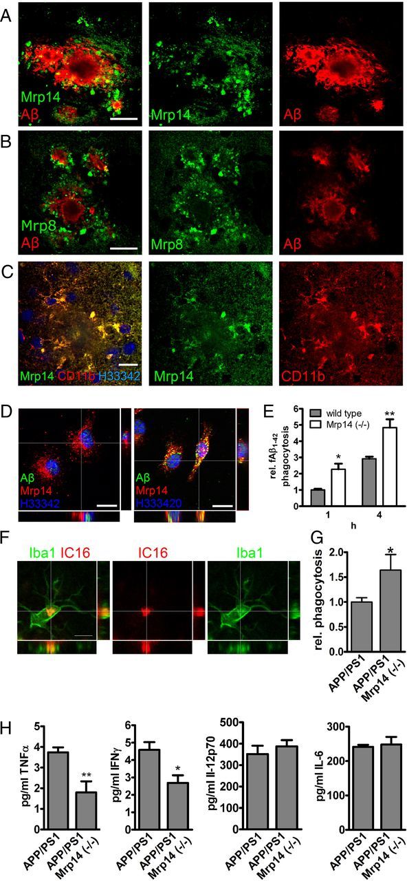
A–C, Brain section of 9-month-old APP/PS1 mice were double-stained with antibody IC16 against Aβ and a polyclonal serum against Mrp14 (scale bar, 20 μm) (A), antibody IC16 against Aβ and a polyclonal serum against Mrp8 (scale bar, 20 μm) (B), and a polyclonal serum against Mrp14 and antibody MCA711 against CD11b (scale bar, 10 μm) (C). D, Immunocytochemical detection of Mrp14 in primary mouse microglia in the absence (left) or presence (right) of 0.5 μm FAM-labeled Aβ1–42 for 1 h (scale bars, 10 μm). E, Quantification of FAM-labeled Aβ1–42 uptake in primary mouse microglia from wild-type and Mrp14(−/−) mice using an plate-based assay (representative experiment performed in pentaduplicate ± SEM, Student's t test, *p < 0.05, **p < 0.01). F, Immunostaining of 9-month-old APP/PS1 mouse using antibody IC16 against Aβ and an anti-Iba1 antiserum (scale bar, 5 μm). G, Quantification of the in vivo phagocytosis assay of 16-month-old APP/PS1 and APP/PS1 Mrp14(−/−) mice (mean of n = 4 for APP/PS1 and n = 3 for APP/PS1 Mrp14(−/−) ± SEM, Student's t test, *p < 0.05). H, Determination of the inflammatory cytokines TNFα, INFγ, IL-6, and IL12 p70 in the lysate of APP/PS1 and APP/PS1 Mrp14(−/−) mice by multiplex ELISA (mean of n = 7 ± SEM, Student's t test, *p < 0.05, **p < 0.01).