
Figure 1.
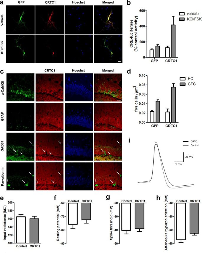
CRTC1 in the DG of the dorsal hippocampus. a, Similar to endogenous CRTC1 protein, plasmid-derived CRTC1 undergoes nuclear translocation following stimulation (KCl/FSK for 4 h) in primary hippocampal neurons. Scale bar, 20 μm. b, CRTC1 increased CRE-dependent transcription following stimulation in primary hippocampal neurons. (CRTC1, n = 4; GFP, n = 4). c, In the DG, CRTC1 is endogenously expressed exclusively in excitatory neurons (not interneurons or glia). Overlap of immunohistochemical staining for endogenous CRTC1 protein (red) and markers of different cell types: green, excitatory neurons (α-CaMKII); glia (GFAP); or interneurons (GAD67 or parvalbumin). Hoechst (blue) identifies DG granule cell layer. Merged images shows that endogenous CRTC1 protein is colocalized in cells positive for α-CaMKII (but not GFAP, GAD67, or parvalbumin). White arrows show lack of CRTC1 staining overlap in GAD67+ or parvalbumin+ cells. Scale bar, 50 μm. d, Context fear conditioning (CFC) increases c-Fos levels in mice microinjected with GFP vector and this effect is potentiated in mice microinjected with CRTC1 vector. CRTC1 overexpression had no effect on c-Fos levels homecage (HC) control mice (CFC–CRTC1, n = 4; CFC–GFP, n = 4; HC–CRTC1, n = 4, HC–GFP, n = 4). e, Input resistance, (f) resting potential, and (g) spike threshold (mV) did not differ between cells infected with CRTC1 vector or control cells whereas AHP (h, i) was decreased in cells infected with CRTC1 vector relative to control cells (CRTC1, n = 7; Control, n = 7). Means ± SEM.