
Figure 4.
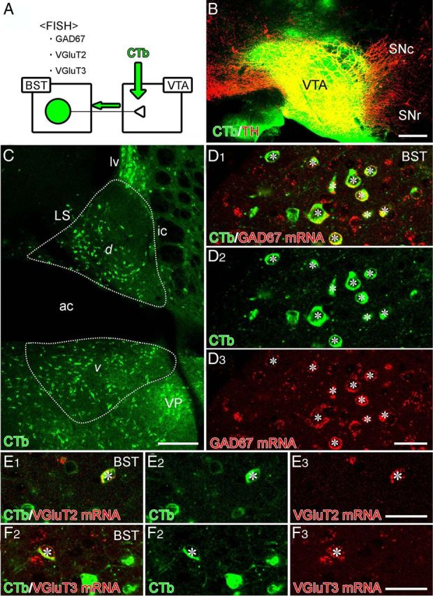
Combined retrograde tracer labeling and fluorescence in situ hybridization (FISH) showing neurochemical composition of BST neurons projecting to the VTA. A, Experimental diagram. B, Injection site of Alexa Fluor 488–CTb (green) in the VTA. This section is immunostained for TH (red) to label DAergic neurons in the VTA and substantia nigra pars compacta (SNc). C, Retrogradely labeled BST neurons projecting to the VTA in dorsal (d) and ventral (v) parts of the anterior BST division. D–F, Double labeling by immunofluorescence for Alexa Fluor 488 (green) and fluorescence in situ hybridization for GAD67 (D), VGluT2 (E), or VGluT3 (F) mRNA (red). Asterisks indicate double-labeled cells. SNr, Substantia nigra pars reticulata. For other abbreviations, see Figure 2. Scale bars: B, C, 200 μm; D–F, 30 μm.