
Figure 7.
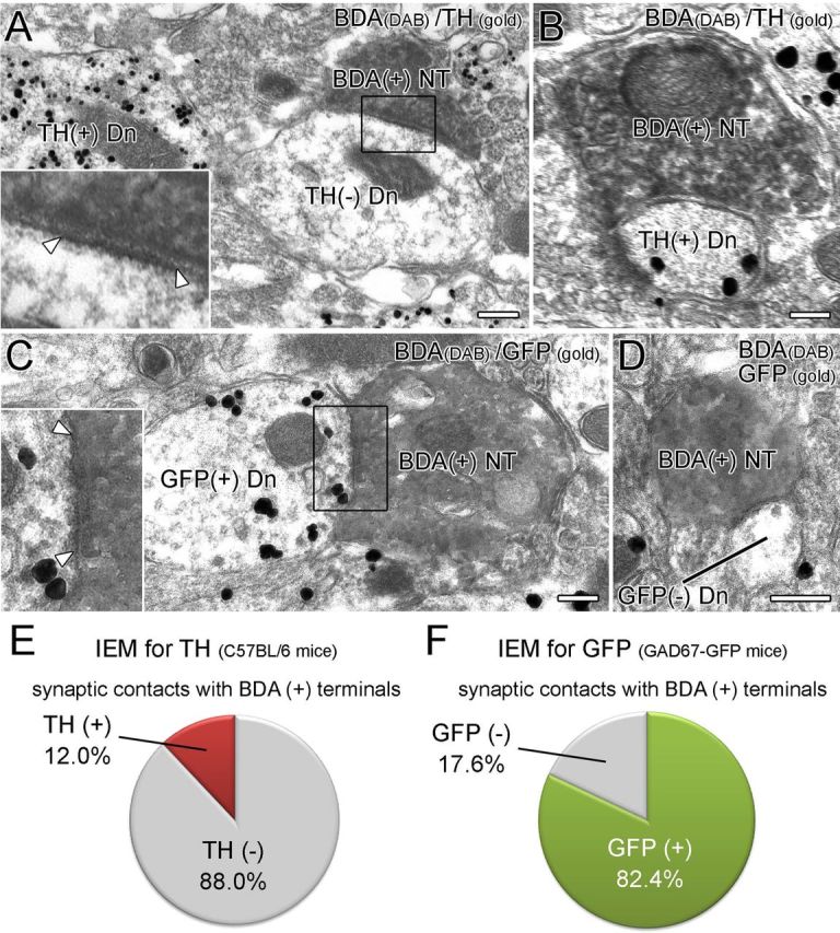
BST neurons forming symmetrical synapses preferentially target GABAergic neurons in the VTA. A, B, Double-labeling immunoelectron microscopy for BDA (diffuse DAB precipitates) and TH (metal particles) in the VTA of wild-type mice. BDA-labeled terminals form symmetrical contact (arrowheads) much more frequently on TH− dendrites (A) than on TH+ dendrites (B). C, D, Double-labeling immunoelectron microscopy (IEM) for BDA (diffuse DAB precipitates) and GFP (metal particles) in the VTA of GAD67–GFP knock-in mice. BDA-positive terminals form symmetrical synapses (arrowheads) much more frequently on GFP+ dendrites (C) than on GFP− dendrites (D). E, F, Proportion of GABAergic (TH− or GFP+) and DAergic (TH+ or GFP−) VTA neurons targeted by BST neurons. Scale bars, 200 nm. Dn, Dendrite; NT, nerve terminals.