
Figure 8.
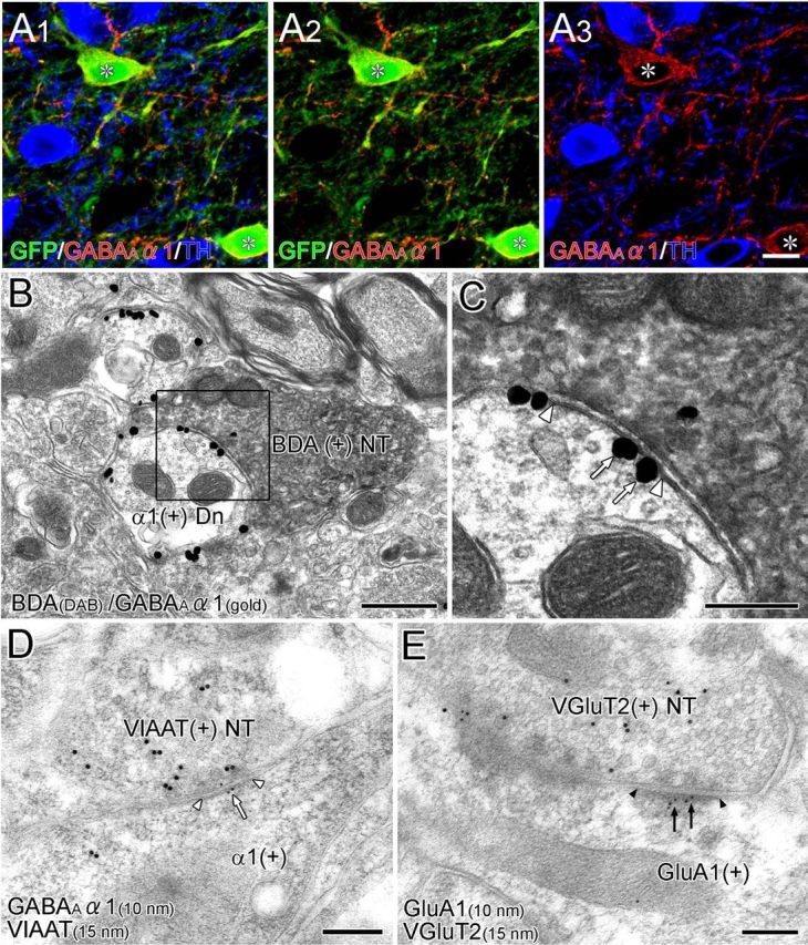
Postsynaptic receptor phenotype in the VTA. A, Triple immunofluorescence for GFP (green), GABAARα1 (red), and TH (blue) in the VTA of GAD67–GFP knock-in mice. GABAARα1 is expressed selectively in GFP-immunopositive GABAergic neurons (asterisk). B, C, Double-labeling preembedding immunoelectron microscopy for BDA (diffuse DAB precipitates) and GABAARα1 (metal particles) in the VTA of wild-type mice. At BST–VTA symmetrical synapses (enlarged in C, white arrowheads), GABAARα1 is localized on the synaptic and extrasynaptic membranes of dendrites (arrows). D, Postembedding immunoelectron microscopy for GABAARα1 (ϕ = 10 nm) and VIAAT (ϕ = 15 nm). VIAAT-labeled terminals form symmetrical synapses (white arrowheads), and GABAARα1 subunit is localized on the postsynaptic membrane (white arrow). E, Postembedding immunoelectron microscopy for GluA1 (ϕ = 10 nm) and VGluT2 (ϕ = 15 nm). VGluT2-labeled terminals form asymmetrical synapses (black arrowheads), and GluA1 is localized on the postsynaptic membrane (black arrows). Scale bars: A, 30 μm; B, 500 nm; C–E, 200 nm. Dn, Dendrite; NT, nerve terminals.