Figure 1.

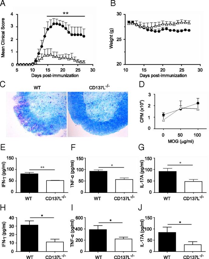
CD137L−/− mice are partially resistant to EAE. A, Clinical score of WT (filled circles) and CD137L−/− (empty triangles) mice immunized with MOG35–55 in CFA and PTx. B, Weight of mice during the course of the disease. Results represent means ± SEM (n = 8–10/group). **p < 0.01. C, Spinal cord sections from EAE mice at day 18 p.i. were evaluated for demyelination using Luxol fast blue staining and cresyl violet for counterstaining. Images are representative staining of 4 mice per group from two independent experiments. D, Lymph node cells from MOG-immunized mice were harvested 10 d p.i. and cultured with indicated concentrations of MOG35–55 peptide for 4 d. Cells were pulsed with [3H]thymidine for the last 18 h of culture and [3H]thymidine incorporation in WT (filled circles) and CD137L−/− (empty triangles) lymph node cells was analyzed. E–G, Levels of IFN-γ (E), TNF-α (F), and IL-17A (G) in the culture supernatants of WT (black bars) and CD137L−/− (white bars) DLN cells cultured in presence of 100 μg/ml MOG peptide were determined by ELISA after 72 h of culture. Results are means ± SEM (n = 3/group). *p < 0.05. H–J, The levels of IFN-γ (H), TNF-α (I), and IL-17A (J) in the sera of WT (black bars) and CD137L−/− (white bars) mice at day 18 post-MOG immunization were determined by BioPlex-ELISA. Results show means ± SEM (n = 3–4/group). Results are representative of three independent experiments.
