Figure 3.
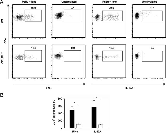
Decreased numbers of effector CD4+ T-cells in CNS of MOG-immunized CD137L−/− mice. Isolated spinal cord cells at day 18 p.i. were restimulated ex vivo with PdBu and ionomycin or media alone for 5 h in the presence of brefeldin-A. Cells were stained for CD45, CD3, CD4 and intracellular IFN-γ and IL-17A expression and analyzed by FACS. A, Dot-plots depict CD4 and intracellular IFN-γ (left) or IL-17A (right) expression derived from WT (top row) and CD137L−/− (bottom row) mice. B, Absolute numbers of CD4+ T-cells producing IFN-γ or IL-17A in spinal cord are shown in the graph. Data represent means ± SEM of 3 independent experiments (n = 3–8) combined for statistical analysis.

