Figure 2.

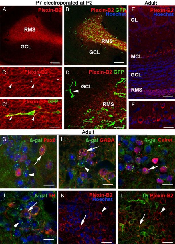
Plexin-B2 is expressed by tangentially migrating neuroblasts and by PG cells. A–D, Sagittal sections at the level of the OB of a P7 mice electroporated at P2 in the SVZ with a GFP-plasmid and immunostained with an anti-Plexin-B2 antibody. Plexin-B2 is highly expressed in the RMS containing tangentially migrating GFP+ neuroblasts. C, C′ is an example of migrating neuroblasts expressing GFP in the cytosol and Plexin-B2 at the cell surface (asterisks). The arrowheads point to the tip of labeled leading processes. Plexin-B2 expression is downregulated in the granular cell layer (GCL), which contains radially migrating GFP+ cells (D, arrowhead). E, F, Sagittal section of the adult OB immunostained with anti-Plexin-B2 and counterstained with Hoechst. Plexin-B2 is detected in the RMS, in the mitral cell layer (MCL), and in the glomerular cell layer (GL), but not in the granule cell layer (GCL). F shows that adult mitral cells are immunoreactive for Plexin-B2. G–L are sections of the adult OB of Plxnb2+/− (G–J) or wild-type mice, at the level of the glomerular layer. In Plxnb2+/− (G–J) OB, β-galactosidase is detected in all the different subtypes of periglomerular cells expressing Pax6 (G, arrow), GABA (H, arrow), calretinin (I, arrow), and TH (J, arrow). Likewise, TH-positive PG cells are immunoreactive for Plexin-B2 (L, arrow). In all panels, the arrowheads indicate cells that express β-gal or Plexin-B2 and are not labeled with the other markers. Scale bars: A, B, 160 μm; C, C′, 22 μm; D, 34 μm; E, 70 μm; F, 30 μm; K, L, 20 μm.
