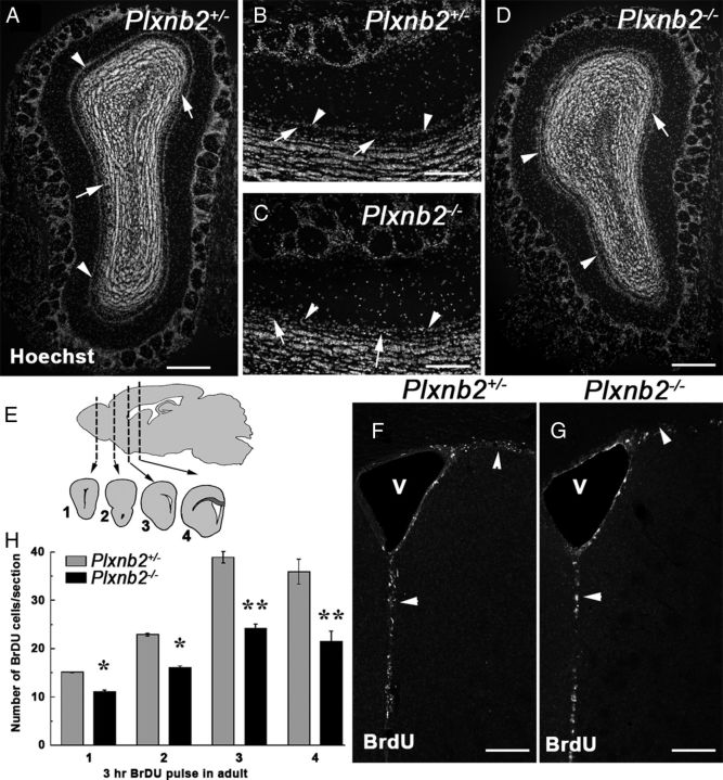
Figure 5.
Perturbed OB layering and proliferation in Plxnb2 mutant. A–D are coronal sections of the OB of adult Plxnb2+/− (A, B) or Plxnb2−/− (C, D) stained with Hoechst. Whereas in the Plxnb2+/− OB (A, B) the mitral cell layer (arrowheads) is well separated from the granule cell layer by the cell-free inner plexiform layer (arrows), in Plxnb2−/− OB (C, D) the two layers are not clearly delineated/distinguishable. E–H illustrate that proliferation is reduced in the adult SVZ of Plxnb2−/− mice. E, BrdU-positive cells (3 h pulse) were counted at four different rostrocaudal levels along the RMS/SVZ as shown on the schematic. F and G are representative pictures of Plxnb2+/− and Plxnb2−/− mice showing BrdU+ cells in the SVZ. H, Quantification of the number of BrdU+ cells per section. A significant reduction of BrdU+ cells is observed in the mutant. Error bars indicate SEM. *p < 0.05; **p < 0.001. Scale bars: A, D, 500 μm; B, C, 250 μm; F, G, 180 μm.

