Figure 6.
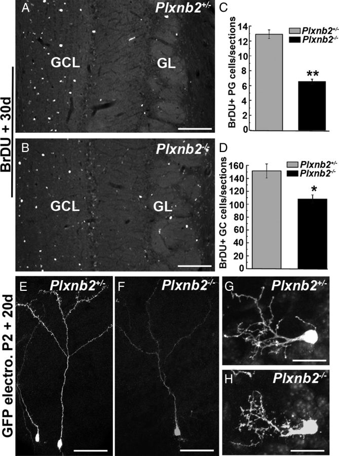
Long-term labeling of newly generated OB neurons in Plxnb2 mutant. A and B are sections of the OB of adult Plxnb2+/− and Plxnb2−/− mice 30 d after an injection of BrdU. In both cases, BrdU+ cells are found in the granule cell layer (GCL) and glomerular cell layer (GL). C, D, The quantification of the number of BrdU+ cells reveals a significant decrease in the number of BrdU+ PG (C) and GC (D) cells in Plxnb2 mutant mice. Error bars indicate SEM. E–H are sagittal section at the level of the OB of P22 Plxnb2+/− and Plxnb2−/− mice electroporated at P2 in the SVZ with a GFP-plasmid. The morphology of GFP+ GC cells (E, F) and GFP+ PG cells (G, H) is similar Plxnb2+/− and Plxnb2−/− mice. Scale bars: A, B, 220 μm; E, F, 300 μm; G, H, 20 μm.

