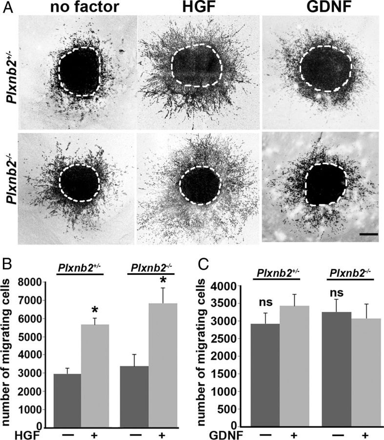
Figure 9.
Cell migration assay and response to HGF and GDNF. A, Confocal image of RMS explants from Plxnb2+/− and Plxnb2−/− P4–P5 mice stained with Topro-3 after 36 h in culture in the absence of any growth factor or in the presence of 50 ng/ml HGF or 200 ng/ml GDNF. B, C, Quantification of the number of cells migrating out of explants cultured with HGF (B) or GDNF (C). No significant difference was observed between wild-type, Plxnb2+/−, and Plxnb2−/− explants cultured with or without growth factors. HGF promoted migration, but GDNF had no effect. Error bars indicate SEM. Scale bar, 200 μm.

