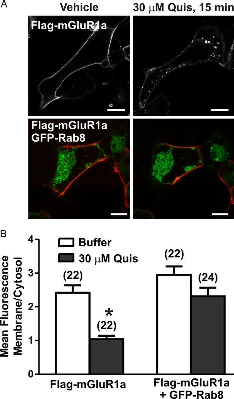
Figure 3.

Live-cell imaging of mGluR1a endocytosis in the absence and presence of Rab8. A, Representative confocal micrograph showing internalization of FLAG-mGluR1a in the presence and absence GFP-Rab8. Live HEK 293 cells expressing 1 μg of plasmid cDNA encoding FLAG-mGluR1a either with (bottom panels) or without (top panels) 2 μg of plasmid cDNA encoding GFP-Rab8 were labeled with Zenon Alexa Fluor 555 on ice, warmed to 37°C, and stimulated with 30 μm Quis for 15 min. B, Quantification of internalization of FLAG-mGluR1a. Data are presented as ratio of membrane fluorescence over intracellular fluorescence and represent mean ± SD of five independent experiments. The number of cells analyzed is provided in brackets in the figure. Scale bars, 5 μm; *p < 0.05.
