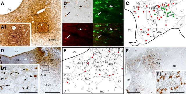
Figure 2.
VLPAG/dDpMe afferents expressing c-FOS during PS-hypersomnia. A, Illustration of a CTb injection site located at the border of the VLPAG and dDpMe in a PS-deprived rat. The enlargement in A1 shows the numerous c-FOS+ neurons within the core of the injection site (delineated by a line) indicating that the neurons specifically active during PS deprivation were effectively targeted. B, Composite photomicrograph illustrating a triple labeling for c-FOS (DAB staining, top left), CTb (in red, bottom left), and MCH (in green, top left) in a PSR animal. An overlay photomicrograph is shown at the bottom right. Arrows point out triple labeled neurons. C, Schematic distribution of CTb/c-FOS/MCH+ (green triangles), CTb/c-FOS+ (red dots), and singly CTb+ (open circles) neurons within the tuberal hypothalamus (AP, −2.6 mm) in a PSR animal. D, Photomicrograph showing that the SLD contains only singly CTb+ (brown cytoplasm) and singly c-FOS+ (black nuclear staining) neurons in PSR animals. The box in D is enlarged in D1. Arrows and arrowheads point to c-FOS+ and CTb+ singly labeled neurons, respectively. E, Schematic illustration of singly CTb+ (open circles), singly c-FOS+ (black dots), and CTb/c-FOS+ (red dots) neurons in the SLD and surrounding structures (AP, −9.2 mm) in a PSR animal. F, Photomicrograph showing in a PSR animal the large number of singly CTb+ cells (brown cytoplasmic labeling) in the Ce after a CTb injection in the VLPAG/dDpMe. The square area is enlarged in F1. Scale bars: A, D, F, 500 μm; A1, D1, F1, 100 μm; B, 200 μm. 3V, Third ventricle; 4N, trochlear nucleus; 4V, fourth ventricle; Aq, aqueduct; BL, basolateral amygdaloid nucleus; CGPn, central gray of the pons; f, fornix; ic, internal capsule; me5, mesencephalic trigeminal tract; mlf, medial longitudinal fasciculus; MPB, medial parabrachial nucleus; mt, mammillothalamic tract; MTu, medial tuberal nucleus; opt, optic tract; VMH, ventromedial hypothalamic nucleus.

