Figure 1.
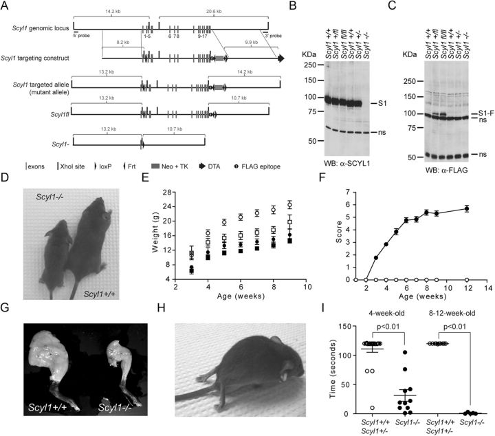
SCYL1 deficiency causes growth defects and early onset progressive motor deficits. A, Schematic representation of the Scyl1 locus, the targeting vector, and predicted Scyl1 mutated locus. After Cre- or Flp-mediated recombination in ES cells, Scyl1− and Scyl1fl loci were generated. Following Cre-mediated recombination, only exon 1 remained. Exons are indicated with gray bars. Black bars represent XhoI sites. Black triangles indicate loxP and Frt sites. Gray box indicates the Neo–TK cassette. Diphtheria toxin cassette is illustrated by a black arrow. The black circle labeled with an f indicates the position of the FLAG epitope. B, C, SCYL1 and FLAG-tagged SCYL1 expression in the cerebrum of Scyl1+/+, Scyl1+/, Scyl1+/fl, Scyl1fl/fl, and Scyl1−/− mice. Cerebrum extracts obtained from adult Scyl1+/+, Scyl1+/−, Scyl1+/fl, Scyl1fl/fl, and Scyl1−/− mice were resolved by SDS-PAGE and analyzed by Western blotting, using antibodies against Scyl1 (α-SCYL1) (B) or the FLAG epitope (C). Note the absence of SCYL1 (S1) in Scyl1-deficient mice. Also note the expression of FLAG-tagged SCYL1 (S1-F) in Scyl1+/fl and Scyl1fl/fl mice. Nonspecific (ns) bands served as loading control. D, Representative photograph of 6-week-old Scyl1+/+ and Scyl1−/− male littermates illustrating the size differences between Scyl1+/+ and Scyl1−/− mice. E, Growth curve of male (circle) and female (square) Scyl1+/+ (white) and Scyl1−/− (black) mice from 3 to 9 weeks of age. Scyl1−/− mice did not recover and stayed smaller than their wild-type littermates. Values are expressed as the mean ± SEM. Males: Scyl1+/+, n = 9; Scyl1−/−, n = 5; females: Scyl1+/+, n = 6; Scyl1−/−, n = 13. F, Disease progression in Scyl1-deficient mice. The clinical progression of defects in mice was assessed by an objective grading system (for details, see Materials and Methods). Values are expressed as the mean ± SEM. Scyl1+/+, n = 13; Scyl1−/−, n = 13. G, Representative photograph of hindlimbs obtained from 12-week-old Scyl1+/+ and Scyl1−/− males showing massive muscle wasting in Scyl1−/− mice. H, Representative photograph of an end-stage Scyl1−/− mouse. The hindlimbs of the Scyl1−/− mouse are atrophied and dorsally contracted. In some mice, flexion of the hindlimb digits is still possible. Also note the flattening of the pelvis. At this stage of the disease, the affected mice move by the power of their front legs. I, Motor defects in Scyl1-deficient mice. The inverted grid test was performed on 4-week-old and 8- to 12-week-old Scyl1+/+ and Scyl1−/− mice to assess their motor functions for up to 120 s as described in Materials and Methods. Four-week-old Scyl1+/+, n = 22; 4-week-old Scyl1−/−, n = 11; 8- to 12-week-old Scyl1+/+, n = 10; 8- to 12-week-old Scyl1−/−, n = 10.

