Figure 3.
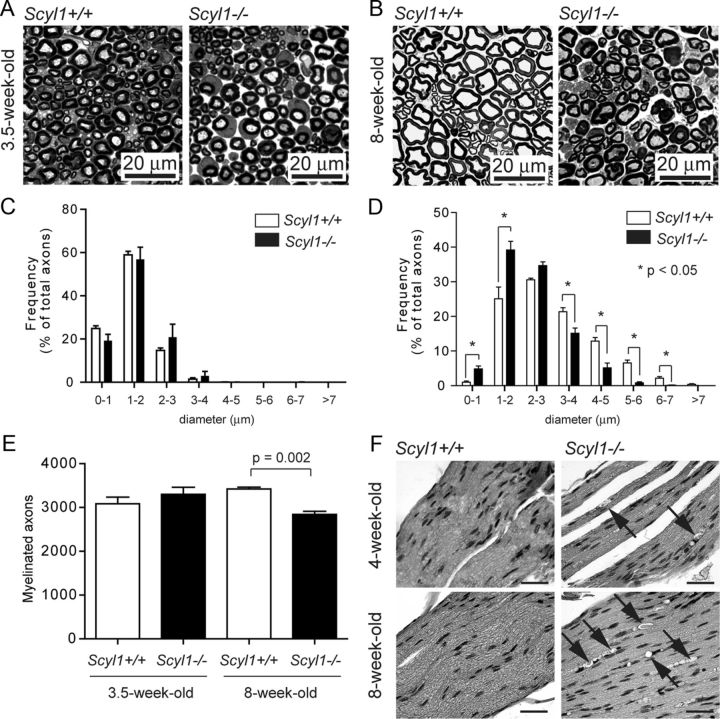
Loss of large-diameter axons in peripheral nerves of Scyl1-deficient mice. A, B, Representative micrograph of toluidine blue-stained semithin sections of the sciatic nerve obtained from 3.5-week-old (A) and 8-week-old (B) Scyl1+/+ and Scyl1−/− mice. Scale bar, 20 μm. C, D, Histogram of the frequency of myelinated axons of the sciatic nerve in function of their axonal diameter in 3.5-week-old (C) or 8-week-old (D) mice. A total of 5882 axons from three 8-week-old Scyl1+/+ mice, 4911 axons from three 8-week-old Scyl1−/− mice, 4050 axons from three 3.5-week-old Scyl1+/+, and 2820 axons from three 3.5-week-old Scyl1−/− mice were evaluated. Note the significant reduction in large-caliber axons (≥5 μm) and increased frequency of small-caliber axons (<1–2 μm) in 8-week-old Scyl1-deficient mice. Values are expressed as the mean ± SEM. *p < 0.05. E, Myelinated axon counts in the sciatic nerve of 3.5- and 8-week-old Scyl1+/+ and Scyl1−/− mice. Values are expressed as the mean ± SEM; n = 3 each. F, Segmental demyelination in the sciatic nerve of Scyl1−/− mice. H&E-stained longitudinal sections of sciatic nerves obtained from 4- and 8-week-old Scyl1+/+ (left) and Scyl1−/− male mice. Arrows indicates myelin ovoids with myelin debris. Scale bar, 50 μm.

