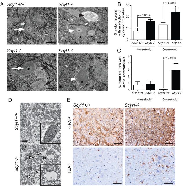
Figure 4.
Degenerating motor neurons in the ventral horn of Scyl1−/− mice. A, Representative micrographs of toluidine blue-stained lumbar ventral horn motor neurons of Scyl1+/+ (top left) and Scyl1−/− (top right and bottom panels) mice. Morphological changes in motor neurons of the lumbar spinal ventral horn included rarefaction of cytosolic organelles, enlarged cytoplasmic vacuoles, and central chromatolysis. White arrows indicate healthy motor neurons. Black arrows point to motor neurons with rarefaction of cytosolic organelles. Black arrowhead points to a motor neuron with enlarged vacuoles. White arrowhead points to a motor neuron showing central chromatolysis. Scale bar, 20 μm. B, Quantification of lumbar ventral horn motor neurons showing rarefaction of cytosolic organelles (including motor neurons with enlarged vacuoles) in 4- and 8-week-old Scyl1+/+ and Scyl1−/− mice. Values are expressed as the mean ± SEM. A minimum of 23 images from three different mice per genotype and age group were analyzed (see Materials and Methods). C, Quantification of chromatolytic lumbar ventral horn motor neurons in 4- and 8-week-old Scyl1+/+ and Scyl1−/− mice. Values are expressed as the mean ± SEM. A minimum of 23 images from three different mice per genotype and age group were analyzed (see Materials and Methods). D, Mitochondrial swelling in motor neurons of Scyl1-deficient mice. In motor neurons of Scyl1+/+ mice, mitochondria of the perikaryon appear normal (top panel and inset). In select LMNs of Scyl1−/− mice, mitochondria are swollen, with disrupted cristae (bottom panel and inset). Mitochondrial swelling was observed in 75 (4.9%) of the 1549 mitochondria analyzed from Scyl1+/+ motor neurons versus 247 (16.7%) of the 1479 mitochondria analyzed in Scyl1−/− mice. Scale bar, 1 μm. E, Neuroinflammation in the spinal ventral horn of Scyl1-deficient mice. Immunohistochemistry using antibodies against Iba1 and GFAP on spinal ventral horn sections obtained from 8-week-old Scyl1+/+ and Scyl1−/− mice. Note the increased Iba1 and GFAP staining in the ventral horn of Scyl1-deficient mice. Scale bars: top row, 100 μm; bottom row, 50 μm.

