
Figure 2.
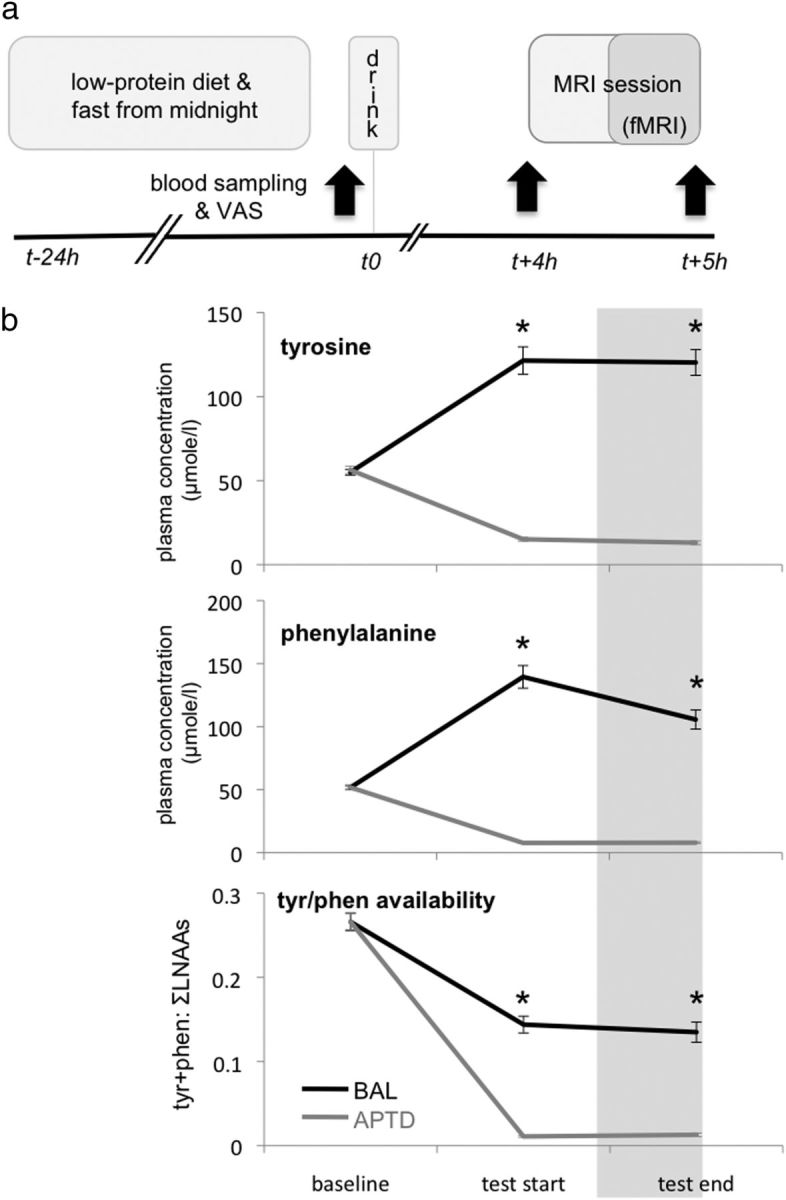
Experimental design and time line. a, The day before testing, participants followed a low-protein diet then fasted from midnight. Upon arrival in the lab, participants completed Visual Analogue Scales (VAS) and a blood sample was drawn. They then consumed an amino acid drink that was either nutritionally balanced (BAL) or deficient in tyrosine and phenylalanine (APTD). VAS and blood samples were repeated (black arrows) 4 h later at “test start” (i.e., just before the MRI session) and again 1 h later at “test end” (i.e., after the MRI session had ended). The first 30 min of the MRI session consisted of installing the participant in the scanner and acquiring anatomical images. The fMRI images were acquired during the last 30 min of the MRI session, ∼4.5 h after initial consumption of the drink. b, Plasma concentrations and availability of tyrosine (tyr) and phenylalanine (phen). At baseline, plasma concentrations of both tyrosine (top) and phenylalanine (middle) were equivalent in the APTD and BAL sessions but then diverged, with significantly (*) lower concentrations at both test start and test end during the APTD session. Brain availability of tyrosine (tyr) and phenylalanine (phen) (bottom), approximated by the ratio of their plasma concentration to that of all other large neutral amino acids (LNAA) (tyr + phen: ∑LNAAs), was also significantly lower at test start and test end during the APTD session. Error bars indicate SEM. Note that error bars for the APTD condition are too small to clearly visualize.