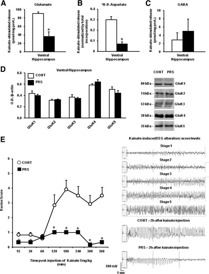
Figure 2.
Reduced kainate-stimulated glutamate release in the ventral hippocampus and kainate-induced limbic motor seizures in PRS rats. Kainate-induced release of glutamate, d-[3H]-aspartate, and GABA in superfused synaptosomes prepared from the ventral hippocampus of control (CONT) or PRS rats are shown in A, B, and C, respectively. Data are expressed as reported in Figure 1, as kainate-induced overflow. Values are means ± SEM of six experiments run in triplicate (3 superfusion chambers for each experimental condition). *p < 0.05 or p < 0.01 versus the respective controls. Immunoblot analysis of GluK1–5 kainate receptor subunits in the ventral hippocampus of control and PRS rats is shown in D. Values are means ± SEM of six determinations. Behavioral score of kainate-induced seizures in control and PRS rats is shown in E. Kainate was injected at the dose of 7 mg/kg, intraperitoneally. Values are means ± SEM of six determinations. *p < 0.01 versus the respective data obtained in control rats. Representative EEG traces corresponding to different stages of kainate-induced seizures and representative traces obtained in control and PRS at 2 h following kainate injection.

