
Figure 2.
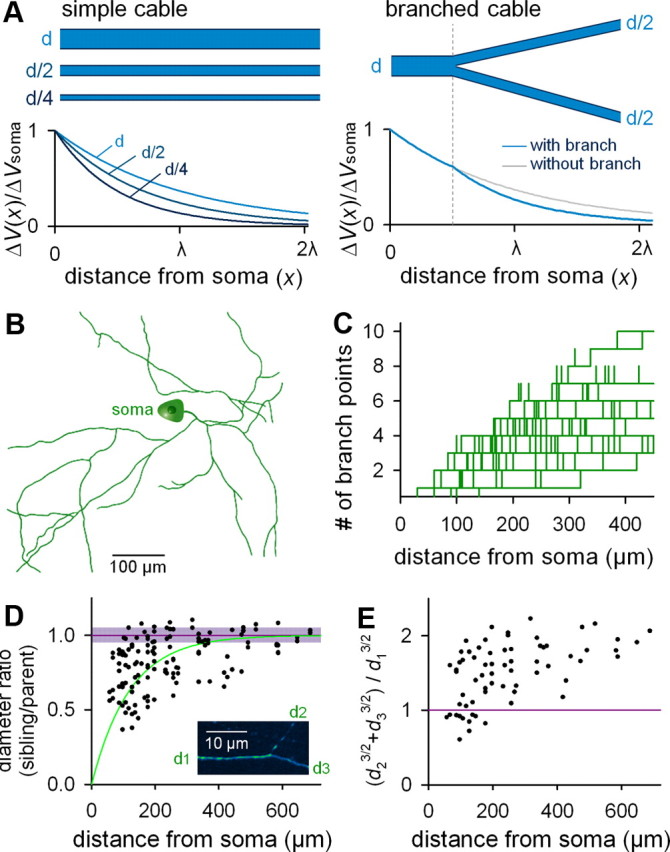
Anatomical characterization of the axonal arborization of CA3 pyramidal cells ex vivo. A, Cable theory predicts that recovery of the depolarized membrane potential (ΔV) along the longitudinal axis of the cable depends on cable diameter (left) and branching (right). B, Representative reconstruction of the CA3 pyramidal cell axon. C, The number of branch points between the soma and a given point on the axon is plotted as a function of the path length from the soma to the examined point (n = 5 cells). D, The ratio of the diameter of a pre-branch “parent” axon segment (d1) to those of the two postbranch “sibling ” axon segments (d2 or d3) is plotted as a function of the path length from the soma. The square root of the Alexa Fluor 488 fluorescence intensity of an axon segment within 10 μm of a given branch point was used as an estimate of axon diameter (n = 5 cells). As control, the mean and SD of the ratio in the fluorescence intensities between two axon points at a path interval of 20 μm without branching are shown in the purple line and shade. Inset, Example of high-magnification of a branching point. E, Ratios of the 3/2 power values of axon diameters [i.e., (d23/2 + d33/2)/d13/2] are plotted as a function of the path length from the soma (n = 5 cells). F, Computational simulation of depolarization-induced AP width along arborized axons when the axonal length constant (λ) is constant throughout the axon (left, without branch decay) or when a branch-dependent decrease in the axonal diameters was considered (right, with branch decay). Simulation was conducted on the neuron reconstructed in B.