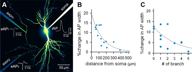
Figure 5.
Depolarization-broadened APs return to a normal width along the length of the axon and through the branch points they traverse. A, Representative image of dual, cell-attached recordings of eAPs from different axonal branches. B, Distance-dependent recovery of depolarization-broadened APs. The curve indicates the least-square best fit of the exponential decay function (Δwidth = 19.6 × e−d/145; d, distance from the soma; F(1,12) = 229.8; p = 0.002). C, Branch number-dependent recovery of depolarization-broadened APs. The curve indicates the best fit of the exponential decay function (Δwidth = 12.4 × e−n/2.9; n, number of upstream branches; F(1,12) = 158.4; p = 0.006). Data presented here were collected from 14 recordings.

