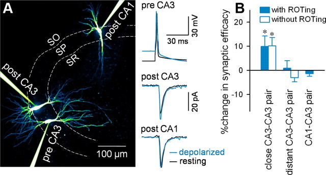
Figure 7.
Somatic depolarization of CA3 pyramidal cells facilitates CA3-to-CA3 but not CA3-to-CA1 synaptic transmission. A, Representative traces of triple recordings from a presynaptic CA3 pyramidal cell and postsynaptic CA3 (close target) and CA1 (distant target) pyramidal cells. Right traces indicate uEPSCs at CA3-to-CA3 (center) and CA3-to-CA1 synapses (bottom) in response to single APs in a presynaptic CA3 neuron (top) in a resting (black) or 20 mV depolarized state (blue). Fifty trials were averaged for each trace. B, Summary graph depicting somatic depolarization-induced changes in synaptic efficacy between CA3-to-CA3 neuron pairs located within 100 μm (close CA3–CA3 pair), CA3-to-CA3 pairs separated by >300 μm (distant CA3–CA3 pair), and CA3-to-CA1 pairs (CA1–CA3 pair). CA3–CA3 neuron pairs were searched with (closed column) and without ROTing (open column). Using ROTing, data were collected from 11 close CA3–CA3 pairs, 13 distant CA3–CA3 pairs, and 12 CA3–CA1 pairs (*p = 0.04, paired t test). Without ROTing, data were collected from five close and four distant CA3–CA3 pairs (*p = 0.03, paired t test).

