Fig. 5.
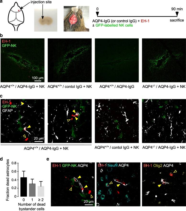
ADCC bystander killing in mouse brain. a Mice were administered AQP4-IgG or control IgG (4 μg) and dead cell stain EH-1 (3 μM) with or without GFP-NK cells (104 cells) by intracerebral injection and sacrificed at 90 min. b Low-magnification confocal microscopy showing dead cells (red EH-1 fluorescence) and green immunostained NK cells. c High-magnification confocal images of AQP4+/+ or AQP4-/- mice treated as in a, showing dead cells (red), GFP-NK cells (green), and astrocytes (white). Yellow filled arrows show dead astrocytes, yellow open arrows show dead bystander cells. d Fraction of dead astrocytes associated with 0, 1 or ≥ 2 dead bystander cells (mean ± S.E.M., 5 slides with > 30 dead cells analyzed). e High-magnification confocal images of mice treated as in a, with indicated stain combinations. Yellow filled arrows show dead astrocytes, yellow open arrows show dead bystander cells some of which were NeuN-positive (neurons) or Olig2-positive (oligodendrocytes)

