Fig. 6.
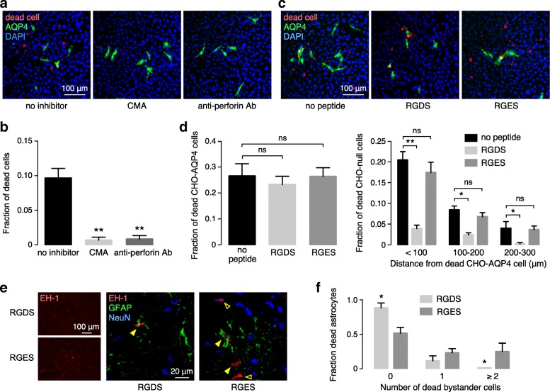
Inhibition of ADCC bystander killing by small molecule, antibody and peptide inhibitors. a CMA and anti-perforin antibody. Cocultures of CHO-AQP4 and CHO-null cell were incubated for 30 min with AQP4-IgG then washed and exposed for 1 h to NK cells with or without CMA (10 nM), and with or without added anti-perforin antibody (10 μg/ml), with fixable dead cell stain added 30 min prior to fixation. b Fraction of dead cells in cocultures from studies as in a (mean ± S.E.M., 4 slides with > 60 dead cells analyzed, ** P<0.01 by unpaired t test). c RGDS peptide. Cocultures were pre-incubated with AQP4-IgG then washed and incubated with RGDS peptide (or control RGES peptide) (200 μM) for 1 h, then exposed to NK cells with dead cell stain added 30 min prior to fixation. d (Left) Cultures treated as in c, showing fraction of dead CHO-AQP4 cells in cocultures with no peptide or RGDS or RGES (mean ± S.E.M., 4 slides with > 30 dead cells analyzed). (Right) Fraction of red-stained, dead CHO-null cells as a function of distance from dead CHO-AQP4 cells (mean ± S.E.M., 4 slides with > 60 dead cells analyzed, ** P<0.01, *P<0.05 comparing no-peptide vs. RGDS or RGES by two-way ANOVA). e Mice were injected with RGDS and RGES peptides (on contralateral side), together with AQP4-IgG (4 μg), NK cells (104 cells) and dead cell stain EH-1 (3 μM). (Left) Low magnification showing EH-1 positive cells. (Right) High magnification confocal images showing dead cells (red), astrocytes (green) and neurons (blue). Yellow filled arrows show dead astrocytes, yellow open arrows show dead bystander cells. f Fraction of dead astrocytes associated with 0, 1 or ≥ 2 dead bystander cells in sections of brains from RGDS and RGES treated mice (mean ± S.E.M., 5 slides with > 30 dead cells analyzed)

