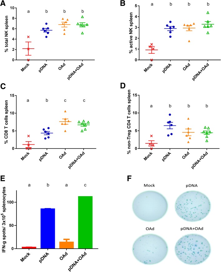
Fig. 4.
Immune cell analysis in the spleen. a Percentage of total NK. b Percentage of active NK. c Percentage of CD8 T cells. d Percentage of non-Treg CD4 T cells. e-f ELISPOT analysis of the splenocytes stimulated with TRP2 peptide. All the results are expressed as mean ± SEM (n = 4–6). a, b and c letters on the graphs indicate significantly different results when the superscript letters are different. The presence of two different letters in two groups indicate a statistical difference (p < 0.05) between them; the same letter in two different groups indicates the absence of a statistical difference between these two groups

