Fig. 3.
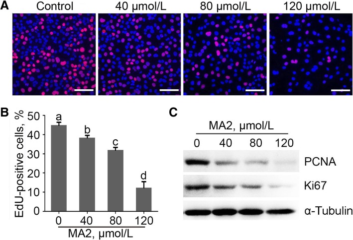
Effect of MA2 on cell proliferation. a GC-1 cells were treated with 0, 40, 80 and 120 μmol/L of MA2 for 48 h. EdU assay was performed to analyze cell proliferation. EdU positive cells were indicated by red fluorescence. Nuclei were stained by DAPI (blue), bar = 50 μm. b EdU positive rates were showed in column diagram. Data were represented by the mean ± SEM, n = 3. Different lower-case letters denoted significant differences (P < 0.05). c Effects of MA2 on expression of the proliferation markers. GC-1 cells were treated with different concentration of MA2 for 48 h. Expression of PCNA and Ki67 were detected by western blot. α-Tubulin was used as the loading control

