Fig. 4.
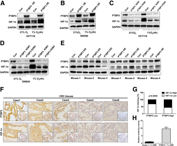
PTBP3 increases HIF-1α protein levels in colon cancer cells. a-b Western blot of HIF-1α expression in HCT116 and SW480 cells ± PTBP3 OE grown under normoxia or hypoxia (1% O2) for 4 h. GAPDH was used as a loading control. c-d, Western blot of HIF-1α expression in HCT116 and SW480 cells ± shRNA PTBP3 KD grown under normoxia or hypoxia (1% O2) for 4 h. GAPDH was used as a loading control. e Western blot of HIF-1α expression in tumor xenografts formed by HCT116 cells ± PTBP3 KD/OE. GAPDH was used as a loading control. f Representative examples of IHC-based correlation between PTBP3 and HIF-1α expression in colorectal tumor sections from TMAs. g Fisher′s exact test analysis of the correlation of PTBP3 and HIF-1α expression in CRC TMAs. h Relative HRE-Luc activity in HCT116 cells ± PTBP3 OE was detected by the Dual Luciferase Reporter Assay System. The Rluc activity was used for normalization. Data are presented as the means ± SD for experiments in triplicate. ***p < 0.001

