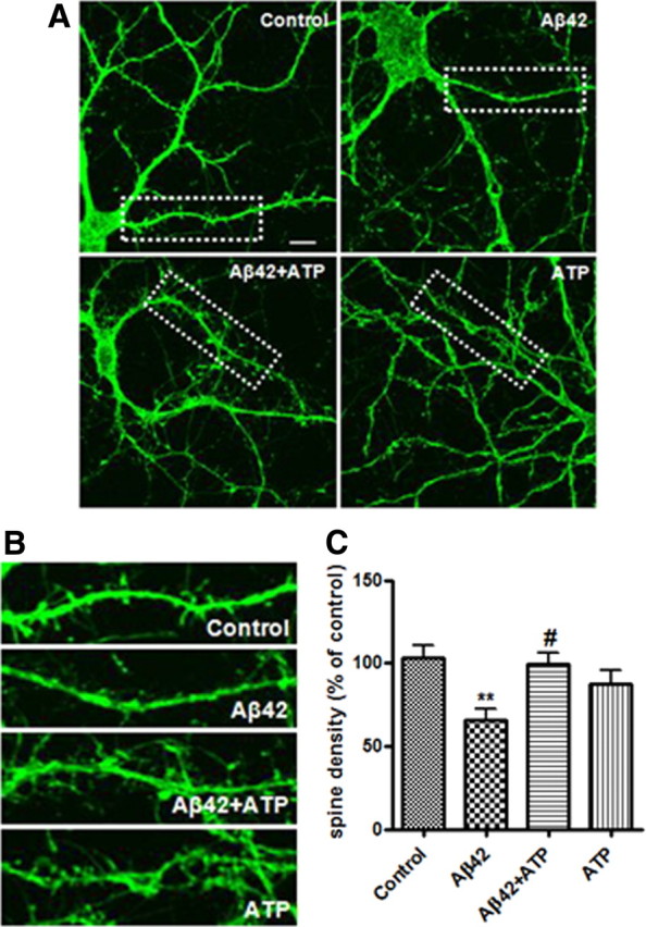
Figure 2.

Effect of ATP on Aβ42-mediated dendritic spine loss in cultured primary rat hippocampal neurons. Primary rat hippocampal neurons (21 DIV) pretreated with or without ATP (10 μm) for 30 min were exposed to Aβ42 (2 μm) for 48 h. A, Rat hippocampal neurons were stained with phalloidin Alexa-488 (green) to visualize dendritic spines. B, Magnified images of the areas marked with dotted lines in Figure 2A. C, Quantification of spine density in dendritic segments. Statistical significance was analyzed by one-way ANOVA followed by a Tukey's multiple-comparison test. **p < 0.01 versus vehicle (control) group; #p < 0.05 versus Aβ42-treated group. Scale bar, 10 μm.
