
Figure 3.
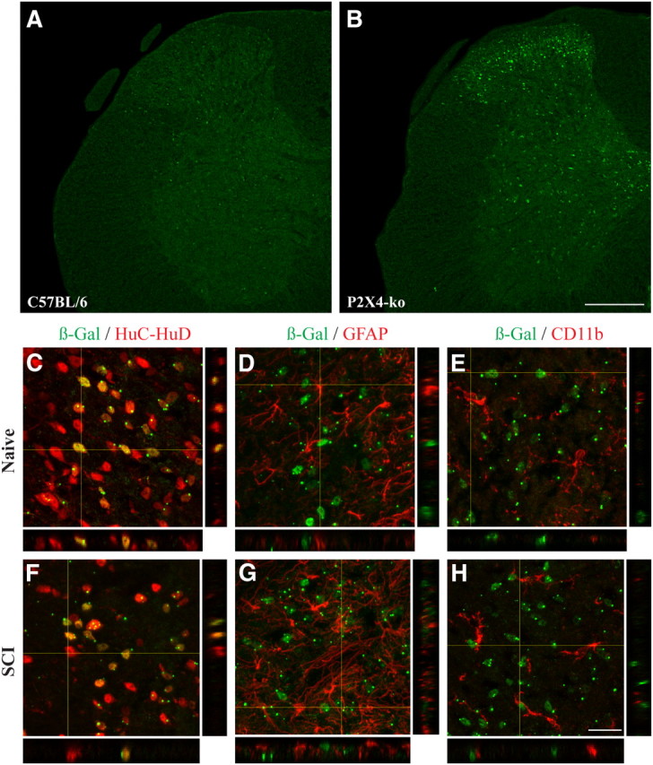
β-Gal staining is localized in neurons in the normal and injured spinal cord of P2X4-KO mice. A, B, Representative confocal photomicrographs showing β-Gal immunostaining (green) in the spinal cord of naive C57BL/6 (negative control) and P2X4-KO mice. Note that, in P2X4-KO mice, the β-Gal reporter gene was inserted in place of the first exon of P2X4, meaning that the P2X4 promoter drives expression of β-Gal in these animals. C–E, β-Gal immunoreactivity colocalized with the neuronal marker HuC/HuD (red, C) but not with GFAP-immunoreactive astrocytes (red, D) and CD11b-immunoreactive microglia (red, E), in the spinal cord dorsal horn of naive P2X4-KO mice. F–H, β-Gal expression colocalized with neurons, but not astrocytes and microglia, at 24 h after SCI. Scale bars: (in B) A, B, 200 μm; (in H) C–H, 20 μm.