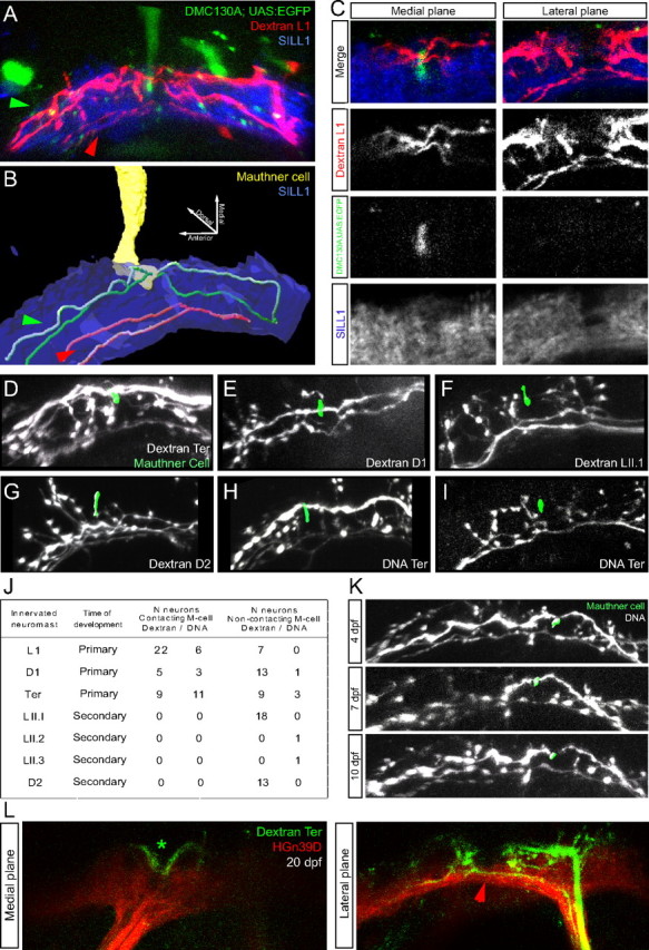
Figure 3.

Neuronal subclassification based on contacts with the Mauthner cell. A–C, Central projections from lateralis afferent neurons of the L1 neuromast (early-born/primary neuromast) labeled by magenta-dextran at 6 dpf, in a Tg[SILL1; hspGFFDMC130A; UAS:EGFP] triple transgenic animal. A is a maximal projection; B is a snapshot of a three-dimensional reconstruction from the data shown in A. C, A detailed view of the central projections toward the Mauthner cell (M-cell) are shown in a medial-to-lateral progression of confocal planes. Green and red arrowheads indicate, respectively, dorsal projections contacting the Mauthner cell and ventrolateral projections non-contacting the Mauthner cell. D–I, Central axons from afferent neurons projecting from early-born/primary neuromasts (D1, terminal) and from late-born/secondary neuromasts (LII.1, D2), labeled by red-dextran (D–G) or by SILL1 DNA injection (H, I), at 6 dpf in a Tg[hspGFFDMC130A; UAS:EGFP] transgenic animals. All pictures are snapshots of the three-dimensional reconstruction of the central projections and the distal tip of the Mauthner cell's lateral dendrite (green). J, Quantification of the contacts between lateralis afferent neurons and the lateral dendrite of the Mauthner cell, sorted by labeling method and neuromast identity: early-born/primary neuromasts (L1, D1, terminal) and late-born/secondary neuromasts (LII.1, LII.2, LII.3, D2). K, Central projections from afferent neurons labeled by SILL1 DNA injection in Tg[hspGFFDMC130A; UAS:EGFP] transgenics, at 4, 7 and 10 dpf. Two lateralis afferent neurons are labeled; one contacts the Mauthner cell whereas the other does not. L, Central axons from afferent neurons projecting from terminal neuromasts and labeled by red-dextran at ∼20 dpf, in the HGn39D transgenic line (colored in red). The lateralis column is shown in red. Medial and lateral focal planes are shown. The asterisk indicates dorsal projections with an indentation. The red arrowhead indicates the ventrolateral projections without the indentation.
