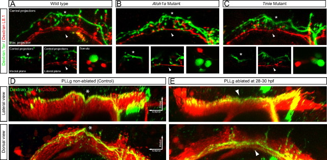
Figure 6.
Analysis of the lateralis neural map in the absence of sensory input and in the absence of early-born neurons. A–C, Central projections and somata from lateralis afferent neurons projecting from the terminal neuromasts (in green, magenta-dextran) and the LII.1 neuromast (in red, red-dextran), in wild-type (A) and homozygous mutants for atoh1a (B) and tmie (C). Neurons were labeled at 6 dpf. Detailed views of both the indentation (asterisk) and the ventrolateral projections (arrowhead) are shown in medial and lateral focal planes, respectively. Top panels in A–C are maximal projections. D, E, Central projections from afferent neurons projecting from the terminal neuromasts that were labeled by magenta-dextran in HGn39D transgenics, whose posterior lateralis ganglion (PLLg) was laser ablated or non-ablated (control) at 28–30 hpf. Neurons were labeled at 6 dpf by dextran uptake. Both lateral and dorsal views of the three-dimensional reconstruction of the central projections are shown. The lateralis column is shown in red. The asterisk indicates the indentation in the dorsal projections. The arrowhead indicates lateral projections without the indentation.

