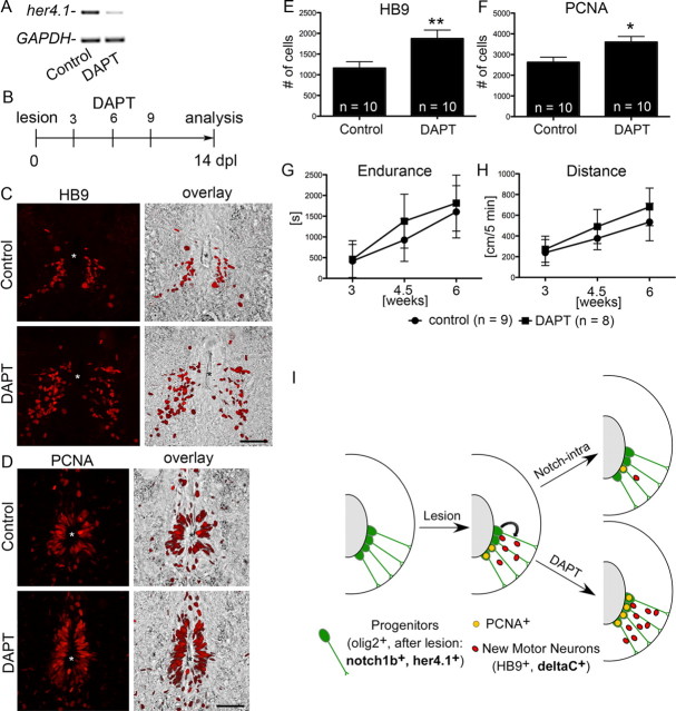
Figure 4.
DAPT treatment increases motor neuron generation and ventricular proliferation and does not impair recovery. Spinal cross sections, centered around the central canal (asterisks), are shown; dorsal is up. A, A single injection of DAPT at 4 dpl reduces her4.1 expression at 5 dpl in PCR. B–F, The number of small HB9+ motor neurons in the ventromedial aspect of the spinal cord (C, E; **p < 0.01) and of PCNA+ cells around the central canal (D, F; *p < 0.05) is increased by the DAPT treatment regimen depicted in B. G, H, Endurance in a flow (G; p < 0.05) and the total distance moved (H; p < 0.01) similarly improved over time for DAPT-treated (as in B but with the indicated time points of analysis) and control fish. I, Schematic summary of results. Overexpression of the intracellular domain of Notch1a attenuates, whereas DAPT treatment augments lesion-induced progenitor cell proliferation (PCNA+) and generation of motor neurons. Scale bars, 50 μm.

