Figure 2.

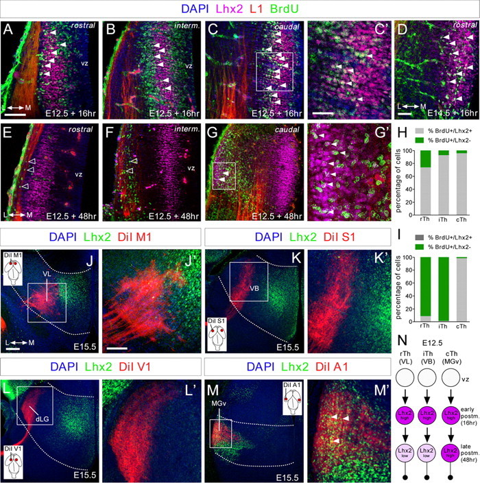
Lhx2 expression is dynamically regulated in postmitotic thalamic neurons. A–G', Coronal sections showing immunohistochemistry for Lhx2 (purple), L1 (red), and BrdU (green), after BrdU was injected at E12.5 and E14.5 (D) and allowed to incorporate for 16 (A–D) or 48 h (E–G'). Nuclear DAPI staining is shown in blue. A large percentage of cells at rostral levels and nearly all at intermediate and caudal levels that incorporated BrdU after a 16 h pulse strongly expressed Lhx2 at E12.5 (A–C', filled arrowheads). D, The majority of the cells that incorporated BrdU after a 16 h pulse strongly express Lhx2 at E14.5. After a 48 h pulse, cells that incorporated BrdU at rostral and intermediate levels showed a strong reduction in Lhx2 expression (E, F, open arrowheads), although it remained high in caudolateral regions (G–G', filled arrowheads). C', G', High-magnification images showing colocalization (white) of Lhx2-positive (purple) and BrdU-positive (green) cells with projecting axons (red) at caudal thalamic levels after a 16 and 48 h pulse, respectively. H, I, Quantification of the percentage of BrdU+/Lhx2+ cells (gray bars) and BrdU+/Lhx2− cells (green bars) along the rostrocaudal axes at E12.5, after 16 and 48 h BrdU injection, respectively. J–M', Coronal sections of DiI-injected brains (red) into M1 (J,J'), S1 (K,K'), V1 (L,L'), and A1 (M,M') cortical areas, showing retrograde-labeled cells in the VL, VB, dLG, and MGv thalamic nuclei, respectively. Immunohistochemistry showing Lhx2 expression pattern (green) at the different thalamic nuclei and its relation with the back-labeled cells from the different cortical areas. Only back-labeled axonal fibers from the A1 cortex showed a strong colocalization with Lhx2-positive cells at caudal thalamic levels (M', arrowheads). Insets show the cortical area in which the DiI crystal was placed. N, Diagram showing the dynamic regulation of Lhx2 protein in thalamocortical neurons. vz, ventricular zone. Scale bars: (in A) A, B, C, E, F, G, 100 μm; (in C') C', D, G', 40 μm; (in J) J, K, L, M, 100 μm; (in J') J', K', L', M', 50 μm.
