Figure 3.

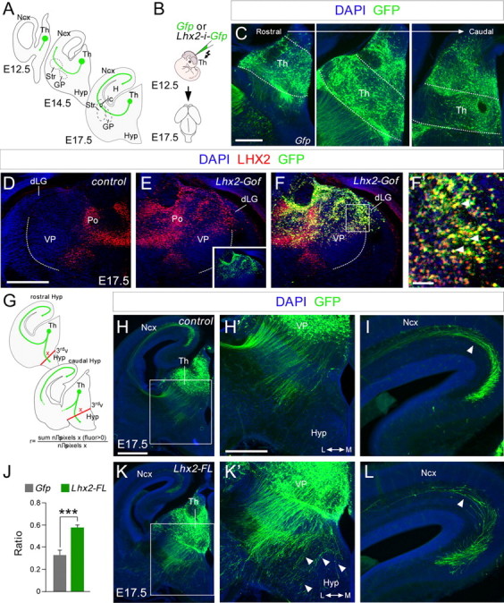
Lhx2 overexpression in the thalamus produces defects in thalamocortical guidance. A, Diagram illustrating how thalamocortical axons travel from the thalamus to the cortex at different embryonic stages. B, Schematic diagram of the experimental design used. C, Example of electroporated brain, showing the GFP expression in coronal sections at rostral, intermediate, and caudal thalamic levels respectively. D–F', Lhx2 immunohistochemistry in control (D) and Lhx2-electroporated (E) hemispheres from the same embryo at E17.5. Note Lhx2 is ectopically overexpressed in the electroporated side (E). We inverted D for a better comparison between the nonelectroporated hemisphere (D) and the electroporated one (E–F'). The inset in E indicates the extension of the electroporated area. G, Diagram of the quantification performed. H–L, Coronal sections of E17.5 brains showing GFP (green) immunohistochemistry after in utero electroporation at E12.5 with Egfp or Lhx2-ires-Egfp-expressing plasmids. Nuclear DAPI staining is shown in blue. H', K', Higher-magnification views of H and K to highlight the thalamocortical guidance defects after Lhx2 overexpression. Derailed thalamocortical axons invade the hypothalamic region (arrowheads) in brains overexpressing Lhx2 but not in control brains. I, L, Fewer electroporated axons reach the neocortex when Lhx2 is upregulated in rTh and iTh neurons (arrowheads). J, Quantification of the derailment of TCAs at the hypothalamus. The ratio is the number of pixels along the red lines showing a positive fluorescence after removing the background fluorescence, divided by the total number of pixels along the red lines. Asterisks indicate significance at ***p < 0.001; Student's t test. Data are presented as the mean ± SEM. Th, thalamus; Ncx, neocortex; Str, striatum; GP, globus palidus; Hyp, hypothalamus; H, hypocampus; 3rdv, third ventricle; Po, posterior group of thalamic nuclei. Scale bars: (in H) H, K, 200 μm; (in C) C, H', I, K', L, 100 μm; (in D) D–F, 100 μm; F', 50 μm.
