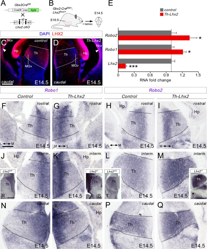
Figure 6.

Conditional deletion of Lhx2 in thalamic neurons augments Robo1 and Robo2 receptor expression in the thalamus. A, B, Schematic diagram illustrating the strategy used to delete Lhx2 in the thalamus at specific developmental stages. C, D, Coronal sections at caudal thalamic levels showing Lhx2 immunohistochemistry in control (C) and in conditional thalamic Lhx2 knock-out mice (D, Th-Lhx2) at E14.5. As expected, Lhx2 expression was strongly reduced in the thalamus of these mice, while the gradient of Lhx2 expression at the cortex remained unaltered (arrowheads). E, Quantitative PCR was performed in the thalamus of control and Th-Lhx2 embryos at E14.5. As expected, the levels of Lhx2 were reduced very significantly in the Th-Lhx2 thalamic embryos. However, the levels of Robo1 and Robo2 were increased compared with the control thalamus. Asterisks indicate significance at *p < 0.05, ***p < 0.001, Student's t test. Data are presented as the mean ± SEM. F–Q, Coronal sections at rostral, intermediate, and caudal thalamic levels showing Robo1 and Robo2 in situ hybridization in control (F, J, N, H, L, P) and Th-Lhx2 (G, K, O, I, M, Q) embryos at E14.5. The expression of both receptors was increased in the absence of Lhx2. R–U, Coronal sections at the caudal thalamic level showing the Robo1 and Robo2 expression in wild-type (R, T) and Lhx2−/− (S, U) embryos at E14.5, respectively. Th, thalamus; Ncx, neocortex; Hp, hippocampus. Scale bars: (in C) C, D, 200 μm; (in F) F–Q, 100 μm; R–U, 200 μm.
