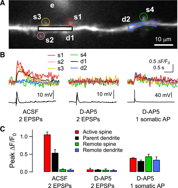
Figure 4.
Activation of multiple spines with focal electrical stimulation near dendritic trees. A, Reconstruction of a distal apical dendrite 180 μm from the soma in a layer 2/3 pyramidal neuron. Electrical stimulation was applied to a glass electrode (e) near the dendrite (10 μm from the dendrite, shown with 50 μm fluorescein). Four spines (s1–s4) and two dendritic regions (d1, d2) were analyzed. B, Two shocks at 40 Hz evoked Ca2+ transients in three spines (s1–s3). Ca2+ transients are also shown in the parent dendrite (d1). In contrast, the remote spine (s4) and dendrite (d2) showed no detectable Ca2+ transients. These Ca2+ transients were blocked by the application of d-AP5 (50 μm), while a single somatic AP evoked dendritic Ca2+ transients in all the spine and dendritic areas. C, Summary plot of the amplitude of Ca2+ transients in spines and dendrites (n = 3 cells). OGB-1 (200 μm) was used as a Ca2+ indicator. An Axioskop 2 FS (fitted with a 40×/0.80 NA objective) equipped with a Noran Odyssey XL was used and the fluorescence signal was captured at a video rate of 25 Hz.

