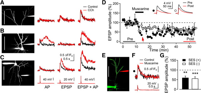
Figure 8.
Dendritic domain-dependent muscarinic induction of CaT2 and LTPm. A–C, A glass stimulating electrode was positioned near dendritic trees in distal apical (A), proximal apical (B), and basal dendrites (C). A somatic AP, synaptic stimulation (EPSP), or EPSP plus AP (10 ms interval) was applied. Left, Reconstructed cells (scale bar, 100 μm) and the stimulating electrodes. Right, representative recordings of the dendritic fluorescence and somatic voltage of nine, five, and nine cells for distal apical, proximal apical, and basal dendrites, respectively. D, Induction of LTDm in distal apical dendrites by muscarinic stimulation. SES (closed circle, indicated by arrow) or stimulation alone at the baseline intensity (open circle) was applied during the application of muscarine. Insets show the average EPSP taken at the time indicated. E, Reconstructed image of a cell (scale bar, 100 μm) and the location of the stimulus electrode (in red). F, Dendritic fluorescence and EPSP evoked by the SES. G, Summary plot of the induction of LTDm with (n = 5) or without (n = 4) the SES in the presence of CCh. **p < 0.01 and ***p < 0.001, respectively.

