
Figure 1.
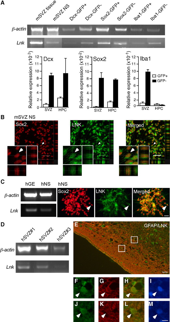
LNK is expressed in SVZ NSPCs in vivo and in vitro. A, RT-PCR and Q-PCR analysis of Lnk expression in tissue and neurospheres derived from mouse SVZ, and in FAC-sorted neuroblasts (Dcx-GFP), NSPCs (Sox2-GFP), and microglia (Iba1-GFP) from SVZ and hippocampus (HPC) of reporter mice. B, Confocal photomicrographs showing LNK immunoreactivity in mouse SVZ neurospheres (NS) costained for SOX2. Double-positive cell depicted by arrowhead on low-magnification images is shown also at higher magnification. C, RT-PCR analysis of LNK expression in GE tissue and NS derived from human fetal GE (hGE). Photomicrographs showing LNK immunoreactivity in human NS (hNS) costained for SOX2. Arrowhead indicates example of double-labeled cell. D, RT-PCR analysis of LNK in three different specimens of adult human SVZ tissue. E, Overview of adult human SVZ stained for LNK (green) and GFAP (red). Boxed areas shown in higher magnification, top box (F–I) and lower box (J–M) depict LNK+ (F, J), GFAP+ (G, K), LNK+/GFAP+ (H, I), and Hoechst+ (I, M) cells. Arrowheads indicate double-labeled cells. Scale bars: B, 50 μm; (in C) C–D, 20 μm; E, 15 μm, and (in M) F–M, 5 μm.