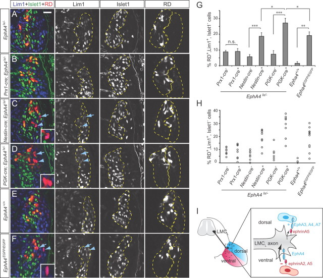
Figure 4.
Guidance errors in EphA4 mutants. A–F, Single confocal plane images of the spinal cord of the indicated genotypes after ventral RD injections and staining for Islet1 and Lim1. Arrows and arrowheads point to Islet1−, Lim1+ LMCL neurons labeled with RD. Examples indicated by arrowheads are shown in the insets at higher magnification. Dashed lines indicate LMCM and LMCL domains. Scale bar: (in A) A–F, 50 μm. G, Quantification of RD-labeled LMCL cells as a percentage of all RD-labeled LMC cells. Numbers of embryos analyzed: Prx1-cre−;EphA4lx/−, n = 8; Prx1-cre+;EphA4lx/−, n = 9; Nestin-cre−;EphA4lx/−, n = 7; Nestin-cre+;EphA4lx/−, n = 8; PGK-cre−;EphA4lx/−, n = 6; PGK-cre+;EphA4lx/−, n = 9; EphA4+/+, n = 3; EphA4EGFP/EGFP, n = 12. Minimum number of RD+ cells counted per embryo: 106. *p < 0.05, **p < 0.01, ***p < 0.001. H, Scatter plots of individual values from each embryo. I, Model of LMCL axon guidance by EphA forward and ephrinA reverse signaling. EphrinAs expressed ventrally act as repellants, triggering forward EphA4 signaling in the axon (thick arrow), while EphAs expressed dorsally act as weak attractants, eliciting reverse ephrinA signaling (thin arrow).

