
Figure 1.
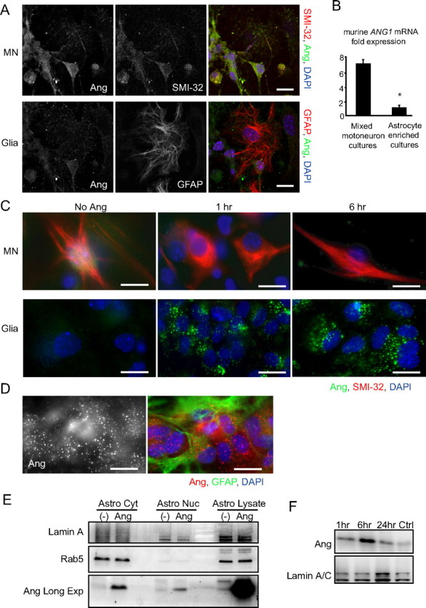
Uptake of recombinant angiogenin appears to be restricted to astroglia. A, Fluorescent images of DIV 7 primary mixed motoneuron cultures costained for SMI-32 or GFAP (red) and Ang (green) imaged from the same field at two different focal planes: neuronal (top) and glial (bottom). B, Quantitative real-time PCR analysis showing murine ang1 transcription levels in primary mixed motoneuron- and astrocyte-enriched cultures relative to β-actin mRNA (*p < 0.05; Student's t test). Data are from n = 3 cultures each. C, Immunocytochemistry of rhAng-treated (1 μg/ml) primary mixed motoneuron cultures costained for SMI-32 (red) and Ang (green). Images show staining in two different focal planes: neuronal (left) and glial (right). D, Representative image of rhAng-treated (1 μg/ml) primary astrocyte cultures costained for GFAP (green) and Ang (red). E, Western blot of rhAng-treated (1 μg/ml) primary astrocyte lysate fractionated into cytosolic (Astro Cyt) and nuclear (Astro Nuc) fractions probed for Lamin A (nuclear marker), Rab5 (cytosolic protein), and Ang [Long exposure blot shown for Ang (Ang Long Exp)]. F, Western blot showing the kinetics of nuclear translocation of rhAng over a 24 h time course uptake in primary astrocytes. Scale bars, 10 μm. All experiments were performed in duplicate with similar results.