
Figure 9.
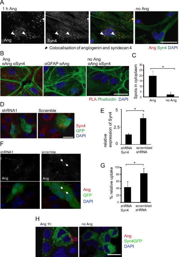
Internalization of rhAng by primary astrocytes depends on syndecan 4 as a receptor. A, Confocal images showing angiogenin (1 μg/ml; 1 h; red) colocalization with syndecan 4 (Syn4; green) staining in primary astrocyte cultures. Experiment was performed in triplicate with similar results, arrowheads indicate colocalization of angiogenin and syndecan 4. B, Confocal images showing PLA of rhAng-treated (1 μg/ml; 1 h) primary astrocytes with anti-angiogenin (αAng) and anti-syndecan 4 (αSyn 4) or anti-GFAP (αGFAP) primary antibodies (phalloidin staining to visualize cell borders), quantification by blob-finder software (C, *p ≤ 0.05; mean ± SD; n > 350 cells in three independent experiments). D, Confocal images of primary astrocytes transfected with syndecan 4 shRNA or scrambled shRNA (transfected cells indicated by GFP fluorescence) and costained with syndecan 4 (red). E, Quantification of shRNA transfection efficiency (p < 0.0005, t test; n = 45 and 56 cells, respectively). F, Confocal images of syndecan 4 shRNA- and scrambled shRNA-transfected (24 h; green) astrocytes treated with rhAng and costained for angiogenin (1 μg/ml; 1 h; red; performed in duplicate with similar results, arrows indicate angiogenin-containing vesicles). G, Quantification of rhAng internalization (1 μg/ml; 1 h; red) by primary astrocytes after shRNA transfection (48 h, *p ≤ 0.05; mean ± SD; n = 3 experiments). H, NSC34 cells overexpressing syndecan 4 (green) can uptake angiogenin (red; performed in duplicate with similar results). Scale bars, 25 μm.