
Figure 7.
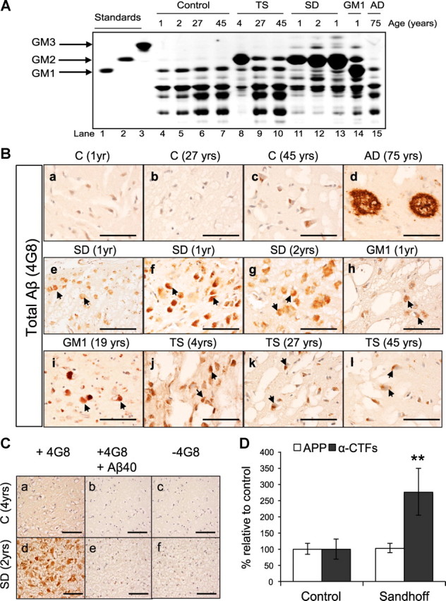
Human Sandhoff brains accumulate intraneuronal 4G8 immunoreactivity and APP-CTFs. A, Thin-layer chromatography analysis of cortices derived from GM1 gangliosidosis (GM1), SD, TS, AD, and age-matched controls, shows the levels of different gangliosides in each brain sample. B, GM1, SD, TS, AD, and age-matched control-derived cortical sections were immunostained using the monoclonal antibody 4G8. The arrows indicate accumulation of intraneuronal APP/Aβ-LIR in 1-year-old (e, f) and 2-year-old (g) SD brains; 1-year-old (h) and 19-year-old (i) GM1 brains; 4-year-old (j), 27-year-old (k), and 45-year-old (l) TS brains. Scale bars: a–l, 200 μm. C, The specificity of 4G8 staining was confirmed by immunostaining cortical sections derived from human SD and control brains with 4G8 antibody preincubated with 100× molar excess of Aβ1–40 peptide (b, e), or with secondary antibody only (c, f). Scale bars: a–f, 200 μm. D, Western blot analysis of seven fibroblast cell lines derived from SD patients or healthy controls show significant accumulation of α-CTFs in the SD fibroblasts (**p = 0.007). Values are the mean of three individual experiments. Error bars indicate SEM.