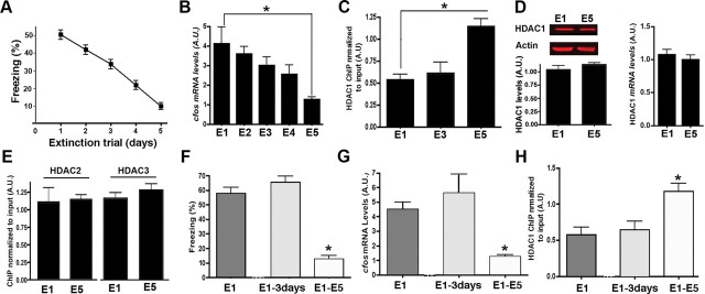
Figure 5.
Fear extinction-dependent recruitment of HDAC1 to the c-Fos promoter. A, Fear extinction in the mice (n = 45) used for molecular analysis in B-E (n = 5/group). B, c-Fos expression was analyzed via qPCR in hippocampal tissue isolated 1 h after exposure to extinction trials. The data are normalized to tissue obtained from a naive control group. C, HDAC1 ChIP was performed from hippocampal tissue 1 h after exposure to E1, E3, and E5. Note that the downregulation of c-Fos correlates with recruitment of HDAC1 to the c-Fos promoter. D, Normalized hippocampal HDAC1 protein levels (left, images show representative immunoblot analysis, 30 μg of hippocampal protein was loaded per lane) and mRNA levels (right) were similar among groups when compared 1 h after E1 and E5 exposure. E, ChIP analysis of the c-Fos promoter was performed for HDAC2 and HDAC3 after E1 and E5. No significant difference among groups was observed. F, Freezing behavior in the E1–3 d group is significantly higher when compared with the E1–E5 group (n = 5/group). G, c-Fos expression was measured 1 h after extinction trials. c-Fos levels were significantly higher in the E1–3 d group when compared with E1–E5 group (n = 5/group). H, HDAC1 ChIP was performed from hippocampal tissue 1 h after exposure to extinction trial in the E1, E1–3 d, and E1–E5 groups (n = 5/group). Note that the increased c-Fos expression in the E1–3 d group correlates with reduced HDAC1 level at the c-Fos promoter. Error bars indicate SEM.

