
Figure 1.
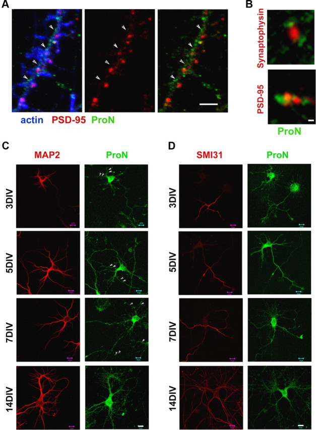
ProN expression during early hippocampal neuron differentiation. A, Primary hippocampal neurons (18 DIV) immunostained for ProN (in green), PSD-95 (in red), and actin (in blue). ProN is localized in close relationship with synapses in mature neurons. The arrowheads indicate that the small puncta of ProN immunoreactivity are in close proximity with PSD-95. B, At higher magnification, localization of ProN (green) and the presynaptic and postsynaptic puncta visualized by synaptophysin (red; top) and PSD95 (red; bottom) labeling, respectively. The close proximity of ProN with presynaptic and postsynaptic puncta can be observed in detail. C, D, Primary hippocampal neurons (3–14 DIV) immunostained for ProN (green) and the dendritic marker MAP2 (in red; C) or the axonal marker SMI31 (in red; D). ProN is expressed in dendrites and axons throughout neuronal differentiation as shown by MAP2 and SMI31, respectively. Scale bars: A, 2 μm; B, 0.1 μm; C, D, 10 μm.